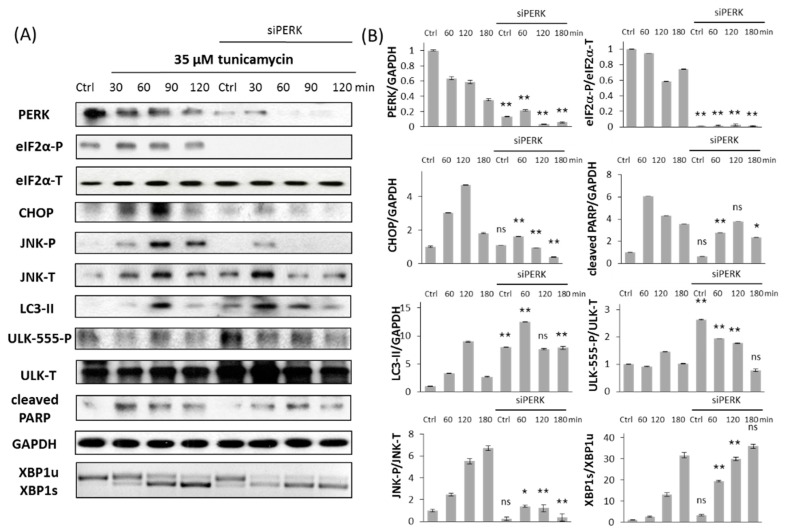
Figure 4.
PERK silencing delays apoptotic cell death at TM treatment. (A) Immunoblot results of key markers. HEK293T cells were treated with 35 µM TM for 3 h without/with use of siPERK. The expression of the crucial autophagy (LC3II, ULK-555-P), apoptosis (cleaved PARP), PERK (PERK, eiF2α-P, CHOP) and IRE-1 (JNK-P, XBP1) markers were followed every 60 min by immunoblotting. All the markers except XBP1 were done by Western blot, while splicing of XBP1 was followed by RT-PCR. GAPDH was used as housekeeping gene. (B) Densitometry data of immunoblotting. The intensity of PERK, LC3II, cleaved PARP, CHOP is normalized for GAPDH, eiF2α-P is normalized for total level of eiF2α, ULK-555-P is normalized for total level of ULK-T, JNK-P is normalized for total level of JNK and spliced XBP1 level is normalized for total level of unspliced XBP1. Three parallel experiments were carried out (errors bars represent standard deviation, asterisks indicate statistically significant difference: * p < 0.05; ** p < 0.01); ns: not significant.

