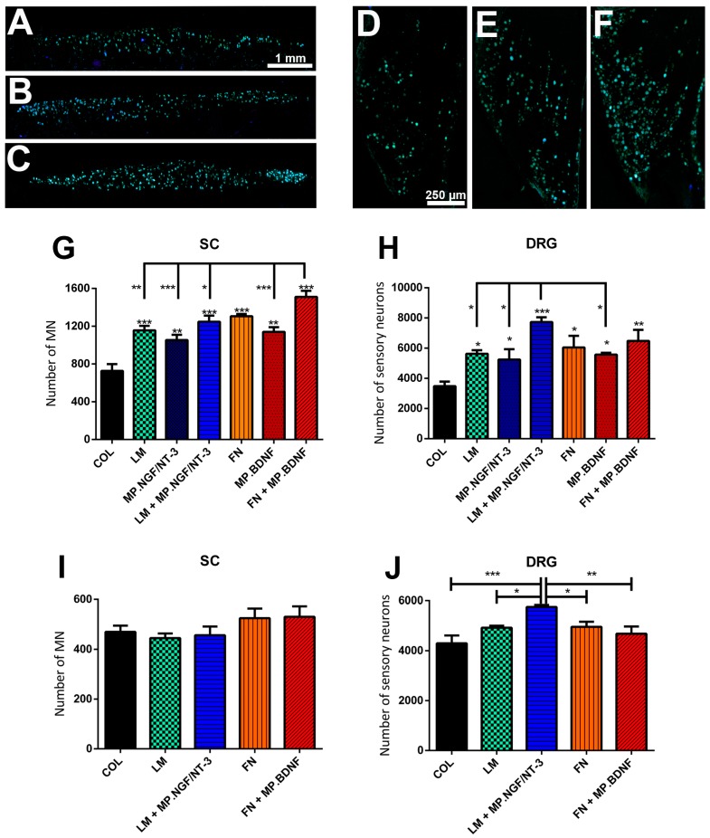
Figure 3.
(A–F) Representative micrographs of neurons retrolabeled with FG in the spinal cord (A–C); and DRG (D–F) of rats after sciatic nerve section and repair with a nerve conduit filled: with COL (A,D); LM + MP.NGF/NT3 (B,E); or FN + MP.BDNF (C,F). (G–H) Histogram of the number of regenerated motor neurons in the spinal cord (G) and sensory neurons in the DRG (H) in the short term (20 days after injury) study. (I,J) Histogram of the number of regenerated motor neurons in the spinal cord (I) and sensory neurons in the DRG (J) after application of FG retrotracer at the ankle level 75 days after injury. Data expressed as mean ± SEM. * p < 0.05, ** p < 0.01 and *** p < 0.001.

