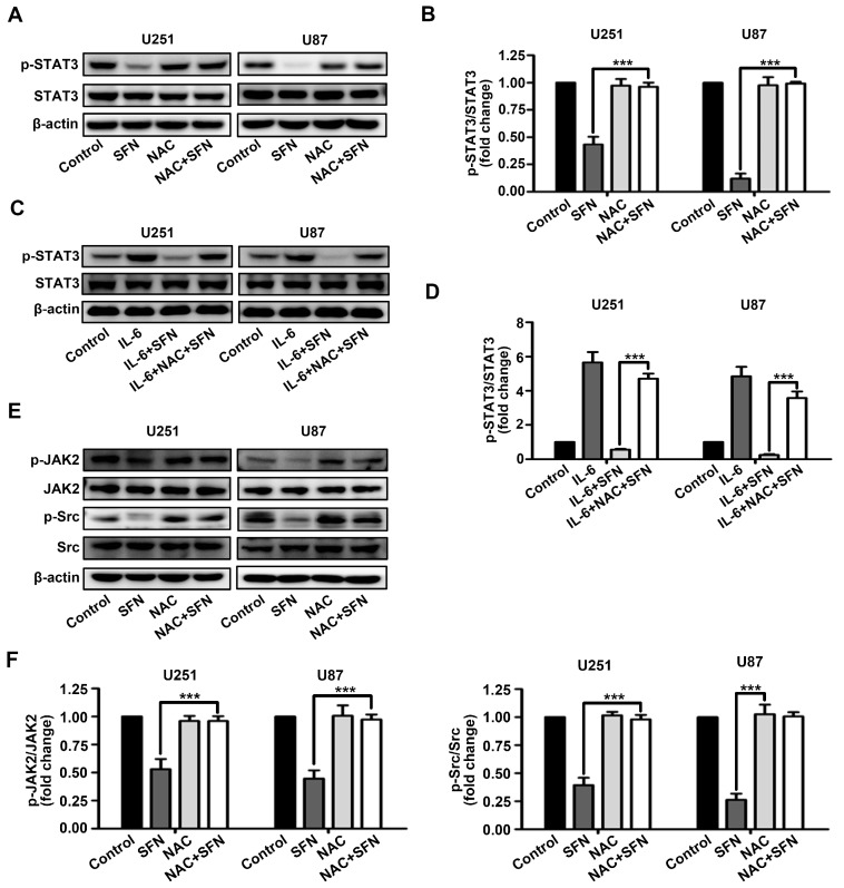
Figure 6.
SFN-induced inactivation of STAT3 signaling in GBM cells is dependent on intracellular ROS generation. (A,B) Cells were treated with SFN (40 µM) for 24 h in absence or presence of NAC. Cell lysates were subjected to western blot to analyze the expression of phospho-STAT3 and STAT3; (C,D) cells were pre-incubated with or without NAC (3 mM), then add IL-6 (50 ng/mL) combined with SFN or DMSO. Cell lysates were subjected to Western blot to analyze the expression of phosphor-STAT3 and STAT3; and (E,F) cells were treated with SFN (40 µM) for 24 h after pre-incubation with or without NAC (3 mM). Cell lysates were subjected to Western blot to analyze the expression of phospho-JAK2, JAK2, phospho-Src, and Src. The bands corresponding to each proteins were quantified and normalized relative to band intensities for control group. Data are represented as means ± SD. *** p < 0.001 versus only SFN or SFN + IL-6 group.

