Figure 1.
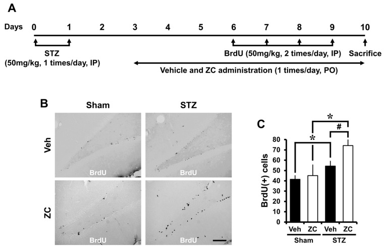
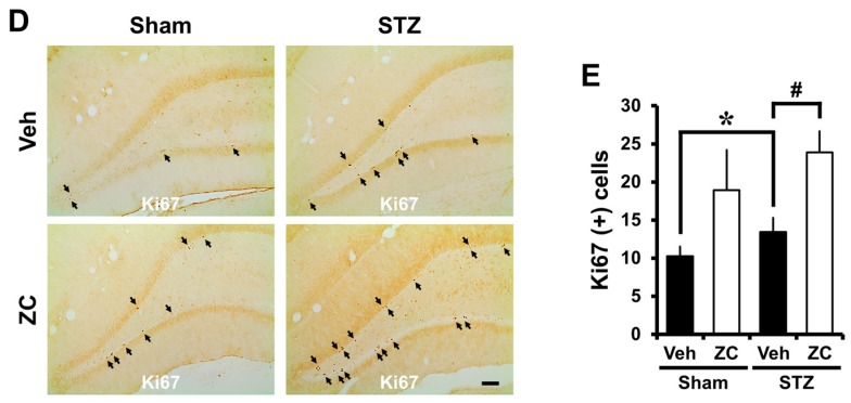
ZC increases proliferation of progenitor cells in the early phase of STZ-induced diabetic rats. (A) Experimental procedure in the early phase of STZ-induced diabetic rats. IP: intraperitoneal, PO: per os; (B) Bright field photomicrographs show BrdU (+) progenitor cells in the hippocampal dentate gyrus (DG). In the early phase, STZ-injected group showed a significant increase of BrdU (+) cells. ZC treatment by gavage for one week after STZ-induced hyperglycemia further increased BrdU (+) cells. Scale bar = 100 µm; (C) Bar graph represents the number of BrdU-positive cells in the subgranular zone (SGZ) of DG. Data are means ± SE, n = 7–12 from each group. * p < 0.05, versus vehicle-treated sham group; # p < 0.05, versus vehicle-treated STZ group; (D) Representative photomicrographs show Ki67 (+) cells in the hippocampal DG. The Ki67 (+) cells is indicated by a black arrow. In the early phase, the STZ-injected group showed a significant increase of Ki67 (+) cells. Short-term ZC treatment increased the number of Ki67 positive cells in STZ-induced diabetic rats. Scale bar = 100 µm; (E) Bar graph represents the number of Ki67 (+) cells in the SGZ of DG. Data are means ± SE, n = 7–12 from each group. * p < 0.05, versus vehicle-treated sham group; # p < 0.05, versus vehicle-treated STZ group.


