Figure 6.
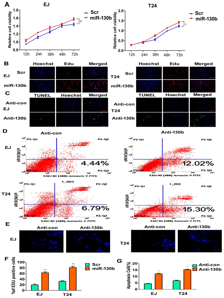
miR-130b-3p affects cell proliferation and apoptosis. (A) CCK8 analysis of EJ or T24 cells transfected with miR-130b-3p mimics or the scramble control; * p < 0.05, ** p < 0.01; (B) Representative images of the Edu assay of EJ or T24 cells transfected with miR-130b-3p mimics or the scramble control. Hoechst stains the nucleus. Scale bar = 100 μm; The blue spots are the nuclear, the red spots are the proliferative cells; (C) Representative photographs of TUNEL staining of EJ or T24 cells transfected with inh-130b or the scramble control. Hoechst stains the nucleus. Scale bar = 100 μm; The blue spots are the nuclear, the green spots are the apoptotic cells; (D) Flow cytometry analysis with Annexin V-PI staining. The percentage of apoptotic cells in EJ or T24 groups transfected with anti-130b or anti-con, respectively; (E) Representative nuclei images of Hoechst staining. Scale bar = 50 μm; (F) Quantitative Edu assay data in five fields. Mean ± SD. ** p < 0.01; and (G) Quantitative Tunel assay data in five fields. Mean ± SD. * p < 0.05.

