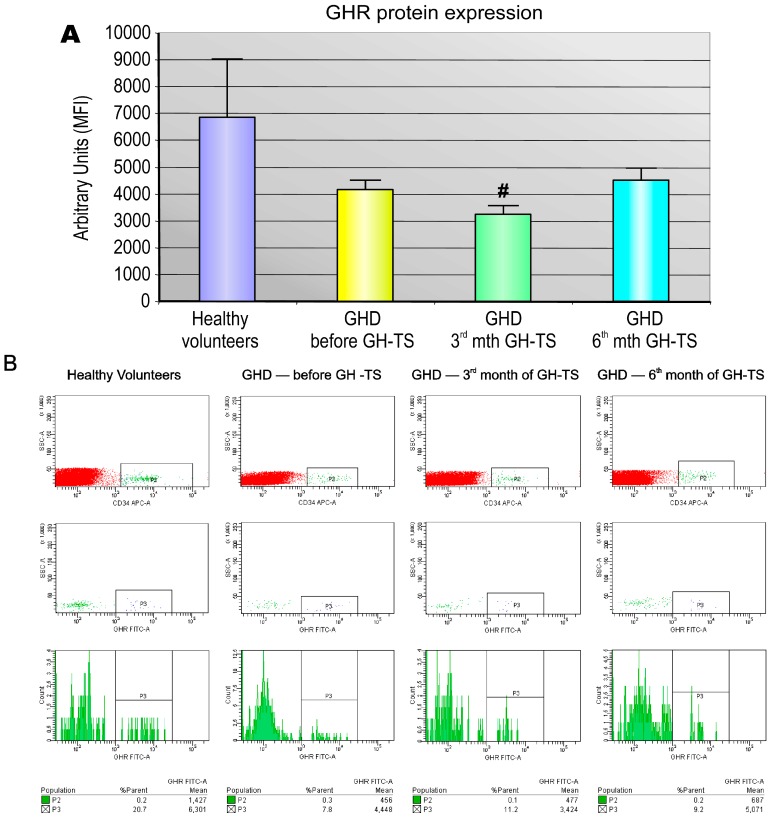
Figure 2.
Quantitative analysis of GHR protein density on CD34+ cells from GHD patients. The quantitative analysis of the expression of GHR receptors on the cell membrane surface of CD34+ cells from GHD patients in course of GH therapy was performed (A). CD34+ cells were collected from PB of from healthy controls and GHD patients at different time points (before GH-TS and in the 3rd and 6th month of GH-TS). Surface GHR protein expression was assessed by flow cytometric analysis based on the mean fluorescence intensity (MFI) of GHR staining of CD34+ cells. The results are expressed as the mean value ± S.D. # p < 0.05 vs. control group. Representative flow cytometric immunofluorescence histograms of GHR expression by CD34+ cells harvested from PB of healthy controls and GHD patients before GH-TS and in the 3rd and 6th month of GH-TS are presented (B).

