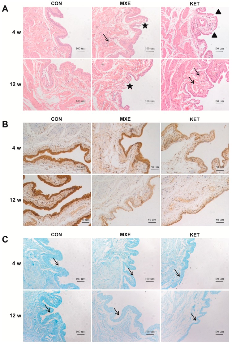
Figure 4.
Mucosal injury in rat bladder tissue after MXE and KET administration. (A) Methoxetamine-treated rats showed mild tissue congestion (arrow) and interrupted continuity of mucosa (asterisks). Magnification: ×100, scale bar: 10 μm. The bladder tissues of ketamine-treated rats were characterized by mucosal laceration (triangles), papillary protrusions, and erythrocyte accumulation (arrows); (B) a decreased staining of E-cadherin was noted in the MXE and KET groups when compared with rats in the control group (magnification: ×200; scale bar: 50 μm); (C) alcian blue staining results. A reduced expression of acid mucopolysaccharide was observed in either MXE or KET groups (arrows, magnification: ×100; Scale bar: 100 μm). The representative images in three treatment groups (n = 6 in each group) were illustrated above.

