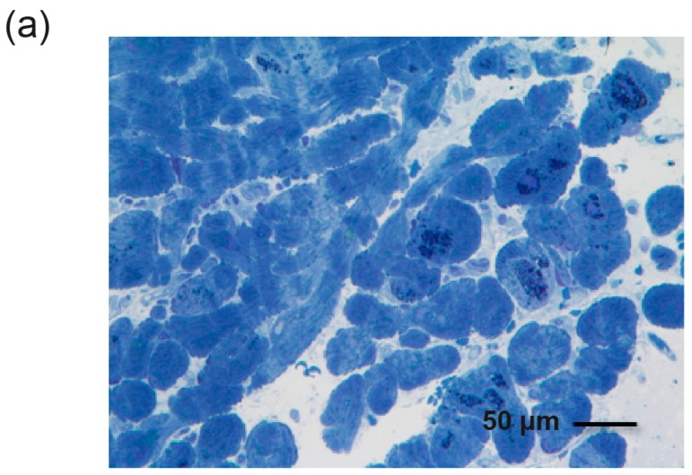
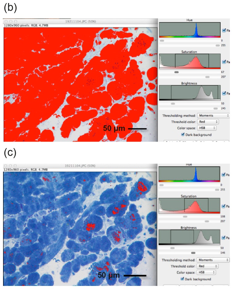
Figure 2.
Image processing using ImageJ software. (a) Cardiomyocyte sample from a single Fabry patient with toluidine blue staining; (b) Total area of the cardiomyocytes (in red); (c) Area of globotriaosylceramide (Gb3) deposition within the cardiomyocytes. The percentage area of Gb3 deposition was calculated from these two values, using ImageJ.


