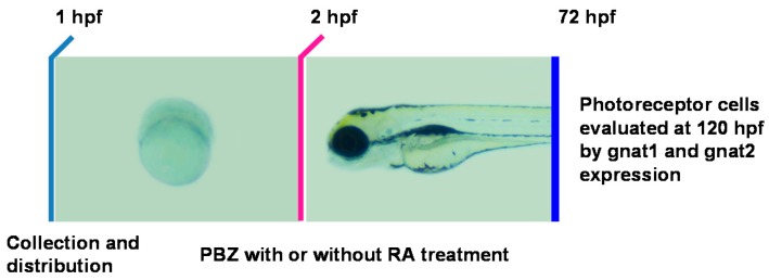
Figure 5.
Schematic diagram showing the timeline of PBZ exposure, RA treatment, and collection of embryos for analysis. Embryos were independently exposed to PBZ (0, 1, or 5 ppm) with or without RA (1 or 5 nM) from 2 hpf until 72 hpf. At 72 hpf, embryos were collected for analysis of retinal photoreceptor cells via gnat1 and gnat2 in situ hybridization.

