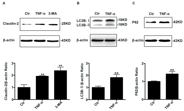
Figure 1.
Change of claudin-2, LC3B-II, and P62 protein in TNF-α treated Caco-2 cell monolayers. (A) Western blotting analysis of caludin-2 in Caco-2 cell monolayer after TNF-α (10 ng/mL) administration or 3-MA (5 mM) treatment for 48 h. (B,C) Western blotting analysis of LC3B-II and P62 in Caco-2 cells after TNF-α (10 ng/mL) administration for 48 h. β-actin was used as an internal control. Data were shown as mean ± SD and replicated three times. ** p < 0.01 versus control group. Ctr, control group.

