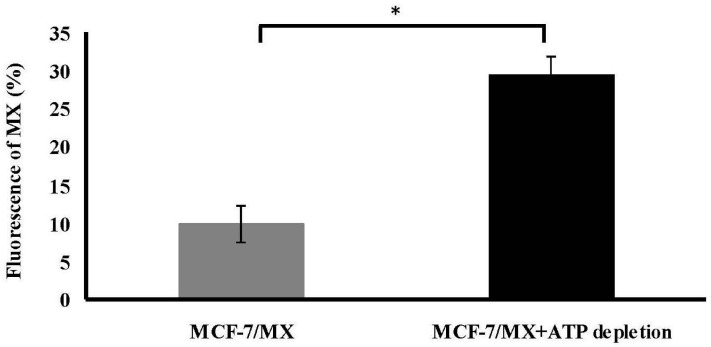
Figure 5.
Treatment of depleted ATP efficiently reduced efflux of MX in MCF-7/MX cells. After ATP being depleted 4 h, including incubation in glucose-free and serum-free medium containing 5 mm sodium azide, MCF-7/MX cells were treated with 10 µL MX for 30 min in complete medium. Fluorescence was measured using the FacsCalibur flow cytometer (Becton–Dickinson). Efflux of MX is significantly decreased in MCF-7/MX cells with 4 h ATP depletion (bar in black). The values represent the mean ± standard deviation obtained from triplicate experiments. * p < 0.05.

