Figure 2.
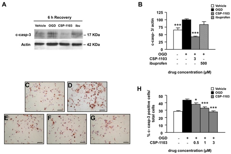
Effect of CSP-1103 and ibuprofen on caspase-3 cleavage in cortical neurons exposed to OGD. (A) Representative images and (B) densitometry analysis of western blot (WB) for c-casp-3 in cytosolic extracts of cells exposed to OGD with or without drugs during 6 h recovery. CSP-1103, but not ibuprofen, was able to reduce c-casp-3 to the basal level. Bars (mean ± SEM) represent the percentage of the casp-3/actin ratio, relative to the OGD value. (C–G) Representative images of immunocytochemistry for c-casp-3 and (H) percentages of c-casp-3-positive cells counted after 24 h recovery ((C) vehicle; (D) OGD; (E) CSP-1103 0.5 μM; (F) CSP-1103 1 μM; and (G) CSP-1103 3 μM). CSP-1103 reduced the number of c-casp-3 immunopositive cells. Bars (mean ± SEM) represent the percentage of c-casp3-positive neurons compared to the total cell number. * p < 0.05, *** p < 0.001 vs. OGD value. +, presence of OGD; −, absence of OGD or treatment.

