Figure 6.
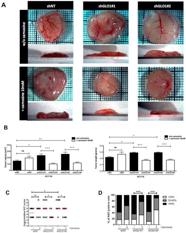
Carnosine treatment of GLO1-depleted HCT116 cells inhibits tumor growth in vivo. HCT116 shGLO1#1 and #2 and shNT control cells were implanted on chorioallantoic (CAM) tumor model. Cancer cells were treated with carnosine (10 mM) from the day after implantation on CAM until the end of the experiment. Tumor growth has been evaluated seven days post-implantation (at least 10 eggs/group) as described under Material and Methods section. (A) Representative macroscopic tumor appearance in each condition is shown; (B) Reduction of tumor volume (left panel) and weight (right panel) after carnosine treatment of GLO1 depleted HCT116-derived tumors, data are shown as mean values ± SEM. Statistical analysis has been performed using Bonferroni Multiple Comparison Test; (C) Significant decrease of argpyrimidine level in experimental CAM tumors upon carnosine treatment. Each dot represents one case and bars represent median. Statistical analysis has been performed using Dunn’s Multiple Comparison Test and Mann–Whitney test; (D) Percentage of Ki67 positive cells in experimental CAM tumors upon carnosine treatment. Statistical analysis has been performed using Chi-square Contingency Test. * p < 0.05, ** p < 0.01, *** p < 0.001, and ns = not significant.

