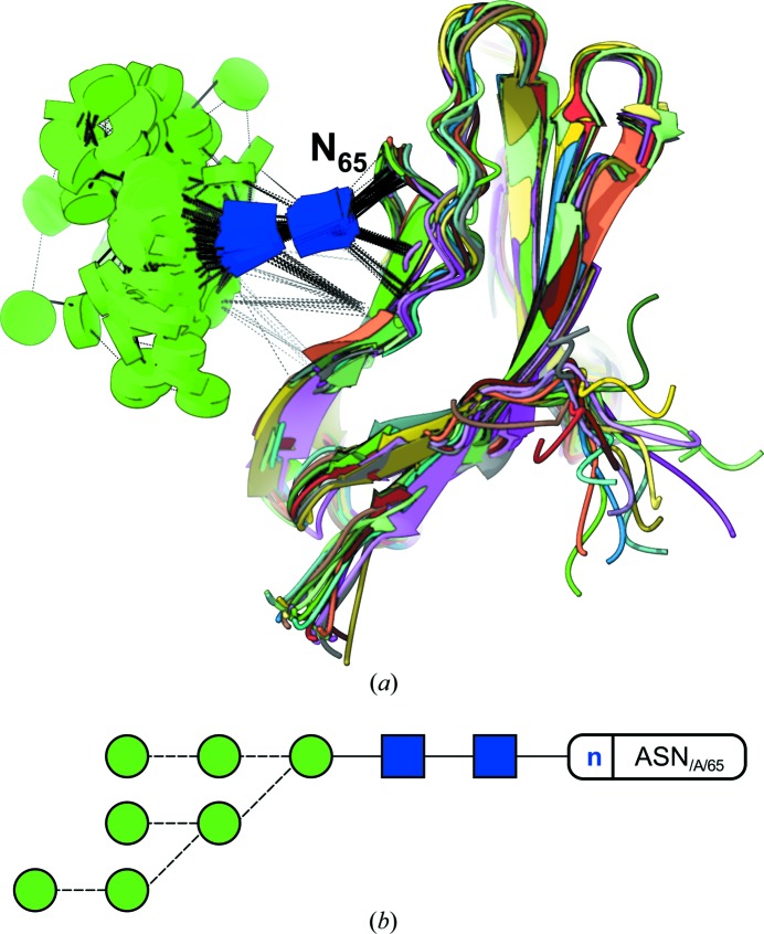
Figure 8.
Simplifying NMR model representation. (a) Glycoblocks view of a partial high-mannose glycan N-linked to the adhesion domain of human CD2. This lateral view of the glycoprotein allows for an unobscured way of looking at the contacts that occur between sugars, and sugars and protein. While hydrogen bonds keep the two core GlcNAc sugars tied to the protein, the rest of the glycan shows great conformational variability. The protein part has been coloured by model. (b) Two-dimensional representation by Privateer. Dashed lines indicate an α-link.

