
Figure 5.
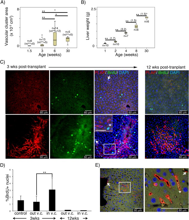
Functional integration of donor‐derived vascular graft in the liver at 3 weeks post‐transplantation. Livers from mice transplanted with E12 FL cells were analyzed at different times post‐transplant. (A): Box plot representing the distribution and mean values of the SCL‐PLAP+ vascular cluster area (v.c.a.) at different ages. The total number of analyzed (n) and v.c. positive (v.c.pos) mice is indicated for each age group. Donor‐derived v.c. were first observed in 3 week‐old mice. The mean v.c.a. values were compared between groups using the U‐Mann–Whitney test (**, p < .05). Significant increment on v.c.a. are observed from 3 to 6 weeks post‐transplant. No significant v.c.a. size increment is observed at long term (from 6 to 30 weeks post‐transplant). (B): Box plot representing the mean values of the liver weight at the different ages. Significant liver growth increment is observed at all ages (Student's t test, **, p < .05) with maximal weight increment from 2 to 4 weeks (fold increment in brackets). Mice were derived from 2 to 3 transplantation experiments for each age. (C): Images from Z stacked optical liver sections from 3 and 12 weeks old FL chimeras, stained with antibodies anti‐PLAP, anti‐BrdU and DAPI. Representative images from liver regions without (top panel) and with (lower panel) SCL‐PLAP+ donor‐derived vascular clusters (v.c.) Most BrdU+ proliferating cells looked‐like hepatocytes (large size, single or multiple round nuclei, prominent nucleoli and moderate to low nucleus/cytoplasm ratio 66), (asterisk, in inset magnified field). Very few BrdU+ endothelial‐like cells are observed (arrows, elongated nuclei, vascular lumen location). (D): Percentage of BrdU+ nuclei referred to the total DAPI+ nuclei determined in regions without v.c. (out v.c.) and within v.c. (in v.c.) as shown in (C). Number of fields to determine average %BrdU+ values: n = 12 from one 3weeks old control mouse; n = 14 from out‐v.c. and n = 10 from in‐v.c., from two 3 weeks old FL chimeras; n = 32 from out‐v.c. and n = 12 from in‐v.c., from two 12 weeks old FL chimeras. **, Unpaired two‐tailed Student's t test assuming p < .05; (E): Optical confocal sections from 3D Z‐stacked confocal images showing PLAP+ endothelial cells forming part of large and micro‐vasculature (arrows) and forming whole functional donor derived vessels containing blood cells (asterisk) connected with circulation. Vessels from 7 out of 10 donor‐derived v.c. presented circulatory cells. Scale bars 60 μm. Abbreviation: PLAP, placental alkaline phosphatase reporter gene.