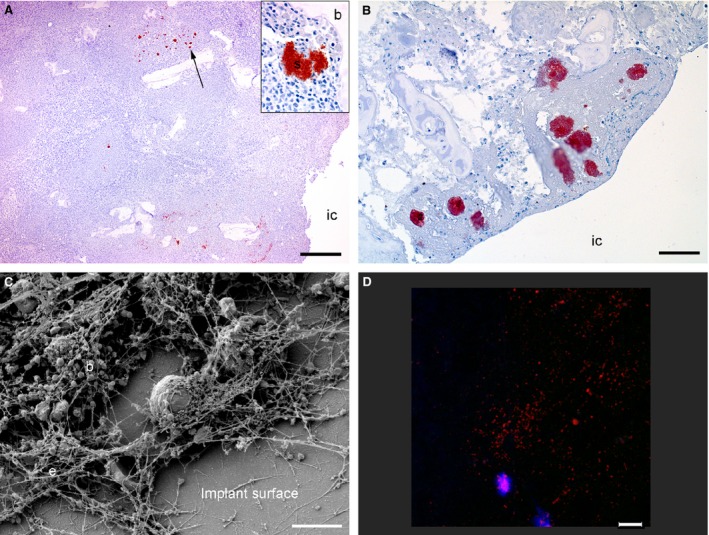
Figure 4.

Visualization of bacteria inside bone tissue and on the surface of steel implants from a porcine model of implant‐associated osteomyelitis inoculated with S. aureus or saline. IC: implant cavity. Picture A: six days after bacterial inoculation, S. aureus‐positive bacteria (arrow) were seen in the peri‐implanted pathological bone area (PIBA). Insert; Close‐up of a positive S. aurous colony, IHC staining for S. aureus, bar = 300. Picture B: four days after inoculation with saline, S. aureus‐positive bacteria were seen enclosed just inside PIBA. These bacteria are supposed to be a result of self‐contamination, as the Spa type of the implant cavity revealed another S. aureus strain as the one used for inoculation, IHC staining for S. aureus, bar = 200. Picture C: scanning electron microscopy (SEM) of an implant surface following 4 days of inoculation with S. aureus showing bacteria (b) and extracellular material (e), bar = 5 μm. Picture D: peptide nucleic acid fluorescence in situ hybridization (PNA FISH) of implant surface following 6 days of inoculation with S. aureus. Bacteria positive for the S. aureus probe light up in red and leukocytes in blue, bar = 20.
