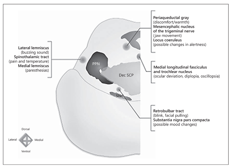
Fig. 2.
Functional mapping of PPN region: correlations between the structures in the vicinity of the PPN and their potential stimulation effects. An electrode positioned lateral to the PPN might be revealed by buzzing sounds (lateral lemniscus), unpleasant painful sensation and/or change in temperature sensation (spinothalamic tract), or paresthesias (medial lemniscus) when trial stimulation is applied. An anteromedial position could elicit the sensation of contralateral facial pulling or blinking (rubrobulbar tract) and/or mood changes (substantia nigra). A mediodorsal location could lead to oscillopsia, diplopia, or ocular deviation toward the side stimulated (medial longitudinal fascicle and trochlear nucleus), mandating careful inspection of extraocular movements. An electrode positioned dorsally could lead to a sensation of discomfort (periaqueductal gray), mandating that the stimulation be done with caution in the vicinity of the PPN. A dorsally placed electrode could also be noticed by jaw movements subjectively felt as pulling of masticatory muscles (mesencephalic nucleus of the trigeminal nerve). A nonspecific altered level of alertness (locus coeruleus) might also be observed. PPN = Pedunculopontine nucleus; Dec SCP = decussation of the superior cerebellar peduncles; SNc = substantia nigra pars compacta. Adapted from Fournier-Gosselin et al. [35], with permission.

