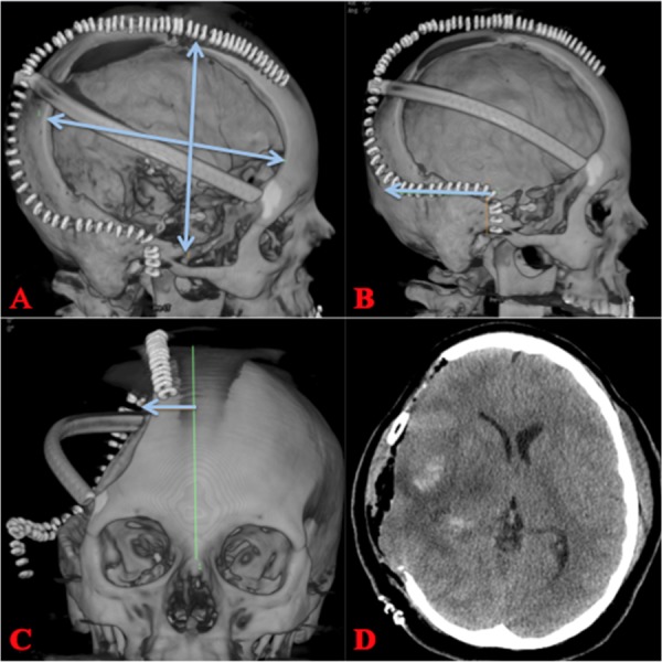
Figure 3. Postoperative Hemicraniectomy.

A, B) A large right hemicraniectomy with dimensions that should be 15 X 12 cm (AP & lateral). The incision should be carried posteriorly for 6 cm from the temporal root of zygoma. C) Incision should be carried to within a couple centimeters from midline to avoid the superior sagittal sinus. D) Postoperative CT showing craniectomy defect and improvement in mass effect.
AP: anteroposterior; CT: computed tomography
