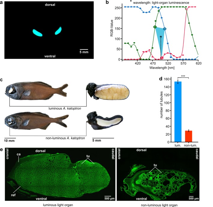
Fig 2. Bioluminescence, anatomical position and structure of the light organs in the splitfin flashlight fish (Anomalops katoptron).
(a) Bioluminescence of A. katoptron during the night. Front view of both subocular light organs. The photograph was taken in a reef tank during the night. (b) Approximated bioluminescence wavelength (498 nm) of the emitted light. (c) Habitus, subocular position and structure of the light organ of the splitfin flashlight fish (Anomalops katoptron) shown for one luminescent and one non-luminescent specimen with degenerated light organs. The oval light organ appears as a white patch because of the guanine crystal reflector on the backside of the light organ and photography using a camera flash. The degenerated non-luminescent light organ illustrates a loss of blood vessels on the surface and a change in shape. (d) Number of tubules in luminous (n = 4) and non-luminous (n = 11) specimens of A. katoptron. Error bars indicate ± SEM (e) Sagittal 3D-photomicrographs of one luminescent and one non-luminescent light organ. The images show the tubules (tu) where A. kataptron host the bioluminescent bacterial symbionts, the reflector (ref), and the cartilaginous light organ attachment (ca) located at the frontal apex.

