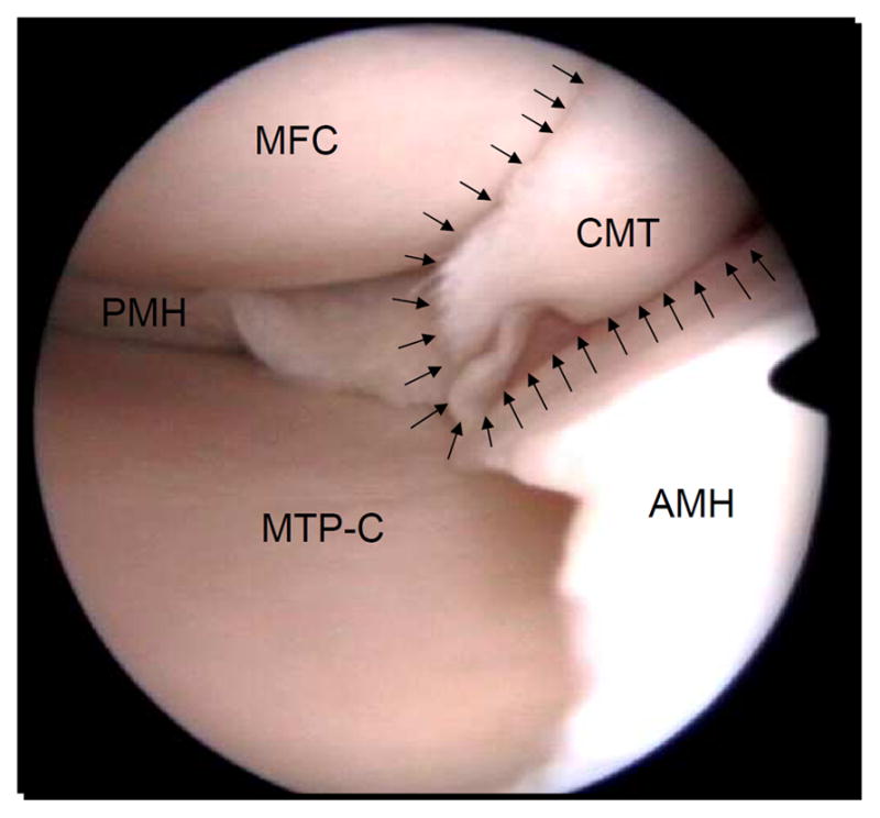
Fig. 3.

Traumatic meniscal tear. Arthroscopic view of a complex traumatic tear of the medial meniscus in a 29-year old man. The round medial femoral condyle (MFC) can be seen in the top left side of the picture, and the corresponding central part of the medial tibial plateau (MTP-C) on the bottom left side of the picture. Note the macroscopic good aspect of the articular cartilage in the medial femorotibial compartment. In the middle right, the complex rupture pattern of the meniscal tear (CMT) located in the pars intermedia can be appreciated (arrows), in part obscuring the view of the medial femoral condyle. The posterior horn of the medial meniscus (PMH) is shown on the left side of the picture and the anterior horn of the medial meniscus (AMH) extends on the right side of the picture.
