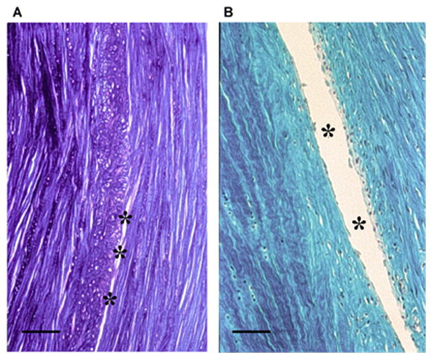
Fig. 5.

Histological evaluation of a complex traumatic canine meniscal tear model. A full-thickness circumferential tear was performed as described in Figure 4. Formalin-fixed, paraffin-embedded sections (5 μm) were taken in the axial plane (12 weeks) and stained with toluidine blue (A) and trichrome (B), highlighting the circumferentially aligned collagen fibers. Evidence of incomplete healing in the tear is demonstrated by the asterisks in the mid-substance region. Scale bar: 500 μm.
