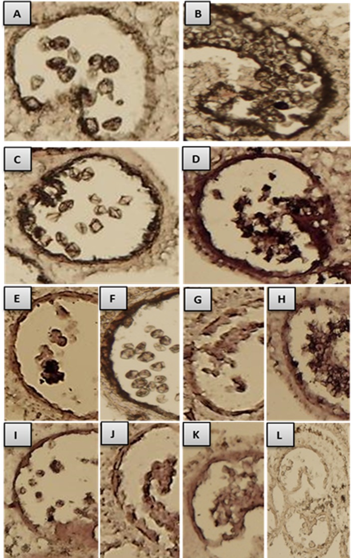
Fig 6. In situ localization of beta-1,3-glucanase, GA2oxs, TA29 and pectinesterase.
A and B: localization of beta-1, 3-glucanase in WT and 7B-1 anthers respectively at binucleate microspores stage. C and D: GA2ox in WT and 7B-1 anthers at binucleate microspores stage, respectively. E and F: TA29 in WT anthers at tetrads and binucleate microspores stages, respectively. G and H: TA29 in 7B-1 anthers at tetrads and arrested binucleate microspores stages, respectively. I, J, K: pectinesterase in WT anthers at tetrads, in 7B-1 anthers at tetrads, and in 7B-1 anthers at arrested binucleate microspores stages, respectively. L: negative control, where a murine miR122a-specific probe was used to ensure that the experimental staining is not an artifact.

