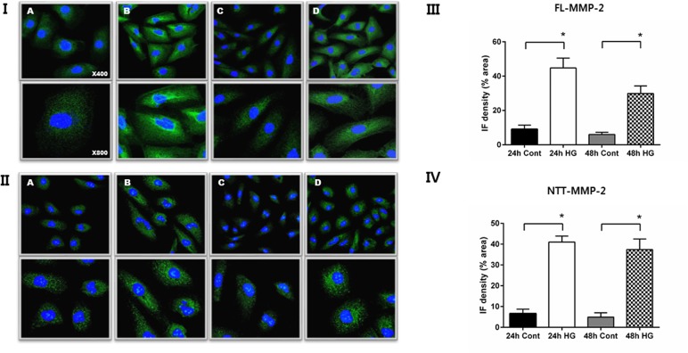
Fig 2. Immunofluorescent (IF) detection of MMP-2 isoform expression in HK2 cells: effect of culture in normal (5 mM) vs. high (30 mM) glucose.
Panel I. IF staining for FL-MMP-2 isoform of HK2 cells cultured in normal (5 mM) glucose for 24 and 48 hours (A, C). IF staining for FL-MMP-2 isoform of HK2 cells cultured in high (30 mM) glucose for 24 and 48 hours (B, D). Upper panel x 400; lower panel x 800. Panel II. IF staining for NTT-MMP-2 isoform of HK2 cells cultured in normal (5 mM) glucose for 24 and 48 hours (A, C). IF staining for NTT-MMP-2 isoform cultured in high (30 mM) glucose for 24 and 48 hours (B, D). Upper panel x 400; lower panel x 800. Panels III and IV: Quantitation of IF staining for the FL-MMP-2 isoform (panel III) and NTT-MMP-2 (panel IV) of HK2 cells cultured in normal (5 mM) and high (30 mM) glucose medium for 24 and 48 hours. (N = 5 for all study groups; *p<0.05).

