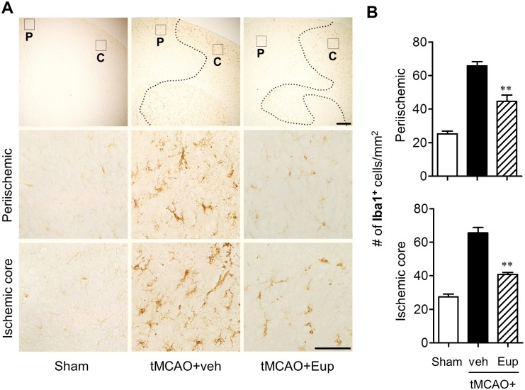
Fig 4. Eupatilin reduces microglial activation in the post-ischemic brain 1 day after tMCAO challenge.
Mice were challenged with tMCAO and eupatilin (Eup, 10 mg/kg, p.o.) was administered immediately after reperfusion. Effects of eupatilin on microglial activation were determined in tMCAO-challenged brain 22 h after reperfusion by immunohistochemistry against Iba1. (A) Representative images for Iba1-immunopositive cells in periischemic (‘P’) and ischemic core (‘C’) regions. Diagram boxes in top panels display brain areas where the images in lower panels were acquired. Dashed lines indicate the lesion site. Scale bars, 200 μm (top panels) and 50 μm (middle and bottom panels). (B) Quantification of Iba1-immunopositive cells in both regions. n = 4 per group. **P<0.01 versus the vehicle-treated tMCAO group (tMCAO+veh).

