
Fig 1. Breakdown of Cal-A into three parts for independent mutagenesis.
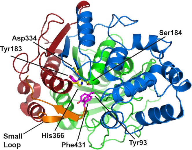
Shown in cartoon representation with the catalytic triad (Ser184, Asp334, His366; yellow sticks) and key residues (Tyr93, Tyr183 and Phe 431; purple sticks). PART 1 (N-terminal region 11–210, in green) is comprised of the α/β fold and includes Tyr93 and Tyr183. PART 2 (tunnel region from 211–350, in blue) has been hypothesized to bind the substrate[25]. PART 3 (from 351 to C-terminal His-tag, in red) contains the small loop (in orange), with Phe 431 that may act as a gate-keeping residue. (PDB identification code: 2VEO)[26]