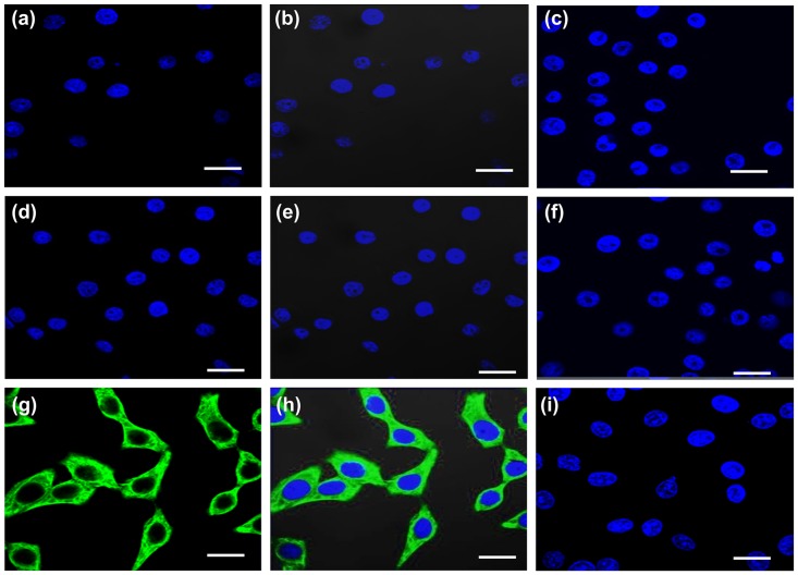
Fig 4. Immunofluorescence localization of vimentin.
hPVECs (a-c) and VK2/E6E7 cells (d-f) did not express vimentin. Cytoplasmic localization of vimentin (green) is seen in HeLa cells (positive control) (g-i). Nucleus was stained with DAPI (blue), FITC (a, d, g), FITC and DAPI merge (b,e,h) and no primary antibody controls (c,f,i) are indicated. The figure shown is one of the representative pictures from three independent experiments (Mag. 63X).

