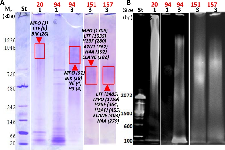
Fig 5. Analysis of DNA-protein complexes in solubilized extracts of four AUP samples using native PAGE.
Non-denaturing 3% acrylamide gels were stained with CBB to visualize proteins (A) and ethidium bromide to visualize DNA (B) in the same gel. Stained areas with high Mr values (protein stain) and large sizes (DNA stain) match and are thus indicative of DNA-protein complexes. These areas were excised, digested with trypsin, and analyzed by LC-MS/MS. Proteins identified are denoted in the gel image with UniProt short names that were also used in Fig 4. BIK is the urinary protein bikunin. Lane and UPsol fraction numbers listed below the sample ID (1 and 3) match. Sample #20 showed DNA release without adding DNase I into the fraction UPsol1. In contrast, the enzyme was responsible for the release of DNA fragments into the fraction UPsol3 for the samples #94, #151, and #157. The lack of DNA solubilization in prior extraction steps is shown for #94 (fraction UPsol1), and proteins in this fraction have generally lower Mr values forming sharper bands, consistent with the absence of association with DNA.

