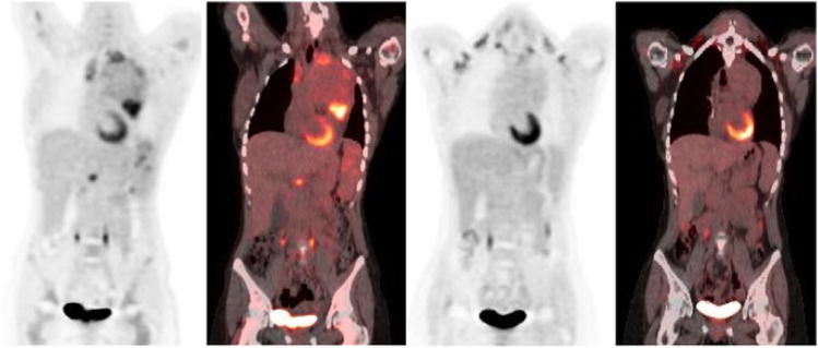
Figure 3.

26 year old female with nodular sclerosis type HL. Baseline PET/CT images (a, b) show large hypermetabolic anterior mediastinal masses, hypermetabolic splenic foci, and periportal adenopathy. Mild marrow hyperplasia was also noted at baseline, bone marrow biopsy was negative. Interim PET/CT (c, d) images show resolution of hypermetabolic activity in the mediastinal mass, spleen and abdominal adenopathy indicating treatment response (Deauville scale 2)
