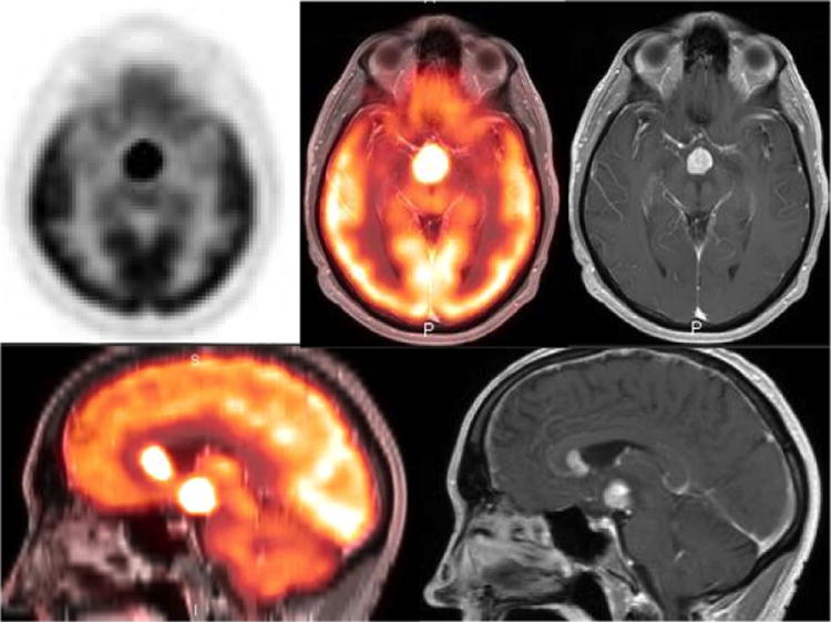
Figure 4.

54 year old male with acute onset headache and confusion. Axial and sagittal PET images of brain fused with contrast enhanced T1 weighted MRI images show an intensely hypermetabolic mass in the suprasellar region. Additional hypermetabolic enhancing nodule is identified in the left frontal horn of lateral ventricle. Histopathology analysis confirmed the diagnosis of large B cell lymphoma.
