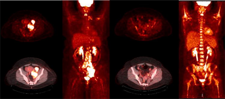
Figure 6.

47 year old female with stage IV-B diffuse large B cell lymphoma B cell lymphoma diffuse nodal involvement. PET and fused PET/CT images show hypermetabolic left iliac adenopathy. The MIP PET image (c) shows additional retroperitoneal, inguinal, mediastinal and left supraclavicular nodal involvement. Interim PET/CT study after 2 cycles of chemotherapy (d-f) shows near complete resolution of adenopathy (Deauville scale 2) with post chemotherapy marrow hyperplasia
