
Figure 9.
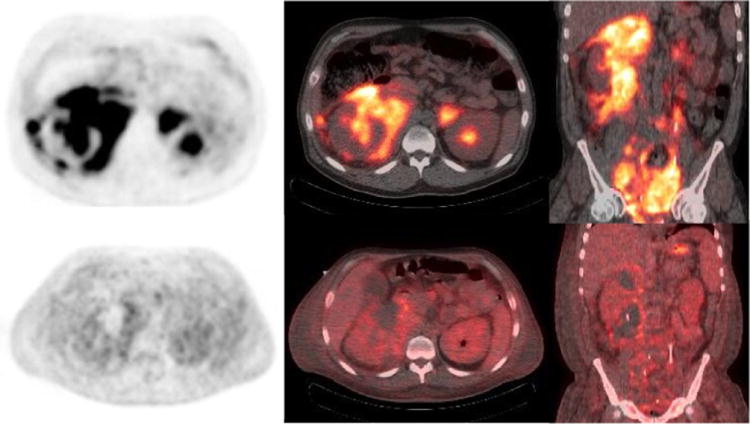
40 year old with Burkitt's lymphoma. Baseline PET/CT (a-c) images show diffuse hypermetabolic abdominal adenopathy with a large perinephric and pelvic hypermetabolic soft tissue mass. Interim PET/CT imaging after 1 cycle of R-CHOP shows significant improvement in metabolic activity of the mass with considerable amount of mildly metabolic residual soft tissue. (Deauville scale 4)