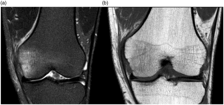
Fig. 1.
MRI at presentation, three weeks after symptom onset. (a) Coronal STIR and (b) coronal T1W images. There was bone marrow edema mostly in the medial half of the medial femoral condyle, otherwise milder and more diffuse in the rest of the condyle, with an appearance more suggestive of contusion than of osteonecrosis. No specific trauma preceded the symptoms, however, the patient had been walking a lot on hard surfaces three weeks earlier.

