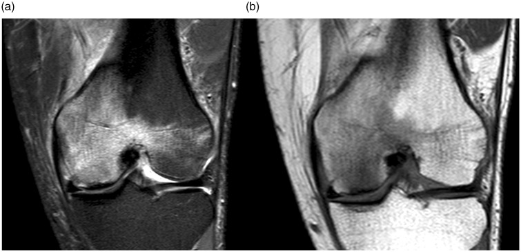
Fig. 2.
MRI three weeks after presentation. (a) Coronal STIR and (b) coronal T1W images. Increase of bone marrow edema both in intensity and extension, with the edema on sagittal images (not shown) now centered around the weight bearing area of the medial femoral condyle, suggestive of osteonecrosis.

