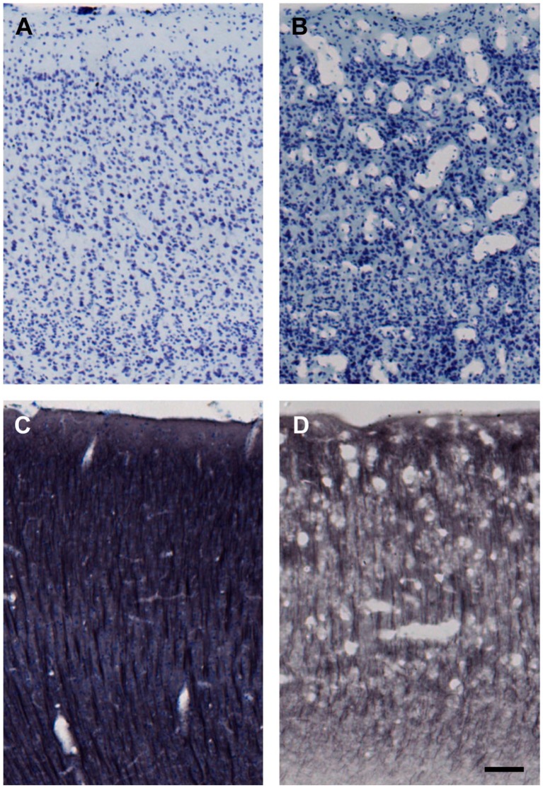
Figure 5.
Freezing artifact produced before cryostorage of sections. All images are from matched locations in the ventral bank of sulcus principalis (cortical area 46). Both brains were cryoprotected using the methods reported here. (A) and (C) are from case AM177, whereas (B) and (D) are from case AM275. Sections in (A) and (B) were Nissl stained right after they were cut from the frozen block (i.e., after step #7 shown in Table 2) and without cryostorage as sections. In contrast, sections shown in (C) and (D) were immunohistochemically processed for microtubule-associated protein 2 (MAP2) with a Nissl counterstain at step #9 (Table 2) after long-term cryostorage in 15% glycerol. Freezing artifact appears as numerous vacuoles that disrupt the laminar cytoarchitecture of the cortex, as shown in case AM275 (B and D). This is likely recrystallization damage and likely occurred during cutting when the brain was inadvertently allowed to warm beyond protocol temperatures (above −30C) at the top of the block (frontal lobe) because ice crystal artifact was not present in occipital lobe sections from the bottom of the same block. Scale bar = 50 µm.

