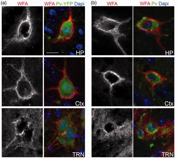
Figure 1.
PNNs ensheath the cell bodies of Parv-expressing neurons in adult mouse brain. (a) WFA-labeled PNN were imaged in hippocampus (HP), cortex (Ctx), and the thalamic reticular nucleus (TRN) of P56 parv-cre::thy1-stop-yfp15 transgenic reporter mice. Parv-expressing interneurons are genetically labeled with YFP in these mice. (b) WFA-labeled PNN were imaged in hippocampus, cortex, and the thalamic reticular nucleus (TRN) of P56 wild-type mice. Parv-expressing interneurons were labeled by IHC. Scale bar = 20 µm.

