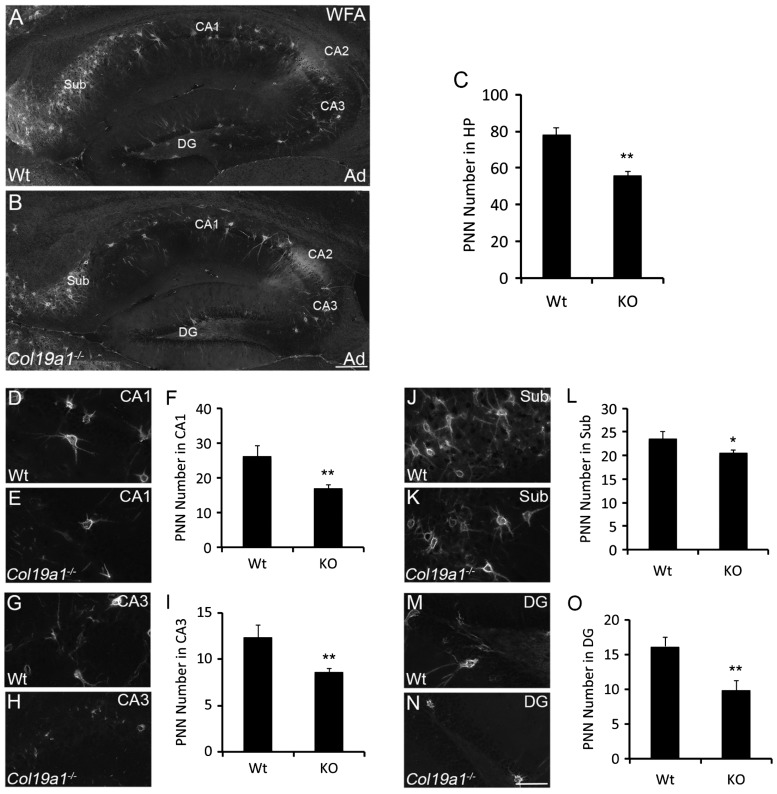
Figure 3.
Loss of WFA-labeled PNNs in collagen XIX-deficient hippocampus. WFA-labeled PNN in hippocampus of P56 wild-type (WT; (a)) and col19a1−/− mutant (KO; (b)). (c) Quantification of WFA-labeled PNN number in P56 WT and KO hippocampus. Data represent means ± standard deviation (SD). ** indicates differs from littermate WT by p < 0.0001 by Student’s t-test. Distribution of WFA-labeled PNNs was assessed in subregions of P56 WT ((d), (g), (j), and (m)) and col19a1−/− mutant ((e), (h), (k), and (n)) hippocampi: Regions of analysis included conru ammonis area (CA) 1 ((d)–(f)), CA3 ((g)–(i)), subiculum (Sub; (j)–(l)), and dentate gyrus (DG; (m)–(o)). Quantification of WFA-labeled PNN number in these subregions are shown in (f), (i), (l), and (o). Data represent means ± SD. * indicates differs from littermate WT by p < 0.05 by Student’s t-test. ** indicates differs from littermate WT by p < 0.0001 by Student’s t-test. Scale bar in (b) = 300 µm for (a) and (b) and in (n) = 50 µm for (d), (e), (g), (h), (j), (k), (m), and (n).

