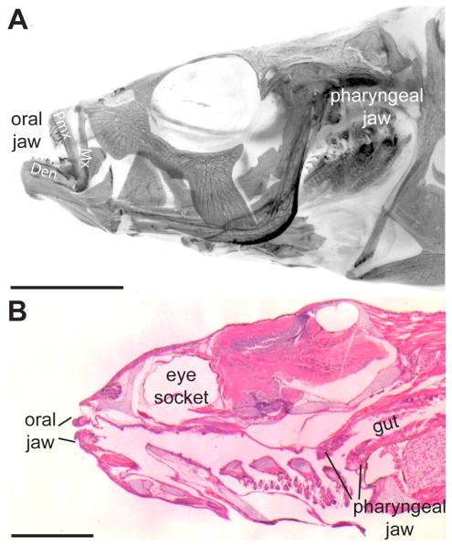
Fig. 1. Stickleback oral and pharyngeal jaws.
(A) Adult 6-month-old freshwater stickleback head (anterior to the left) stained with Alizarin red in whole-mount, imaged under fluorescence (color inverted) after removal of the opercle and subopercle. (B) Hematoxylin and eosin 6 μm sagittal section of 26 days post fertilization marine stickleback. Two sets of toothed jaws are present, the oral jaw in the mouth and the pharyngeal jaw at the back of the branchial skeleton, anterior to the gut. Mx = maxilla, Pmx = premaxilla, Den = dentary bone. Scale bars = 5 mm (A) and 500 μm (B).

