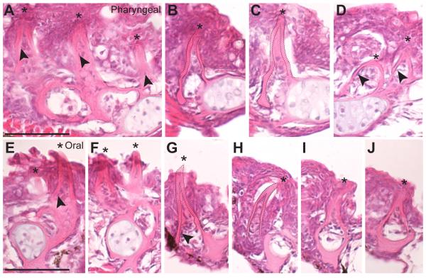
Fig. 3. Oral and pharyngeal teeth are morphologically indistinguishable.
Hematoxylin and eosin stained 6 μm sagittal sections of pharyngeal (A-D) and oral (E-J) teeth on the ventral pharyngeal tooth plate (A-C), dorsal pharyngeal tooth plate 1 (D), dentary (E-F) and premaxilla (G-J). Asterisks mark the tips of teeth, all of which are unicuspid mineralized cones (dashed lines in B,C and G,H). Cells in the pulp cavity likely include presumptive odontoblasts (arrowheads). All sections are from a 26 days post fertilization marine fish. Scale bars = 50 μm.

