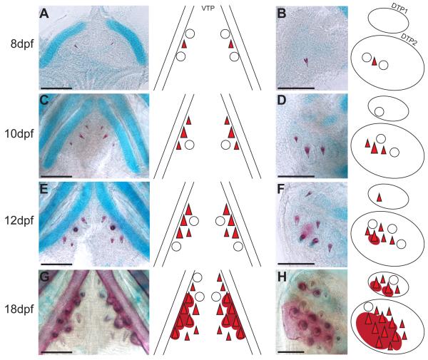
Fig. 5. Developmental time course of pharyngeal tooth formation.
In each panel, bilateral ventral pharyngeal tooth plates (A, C, E, G) or unilateral dorsal pharyngeal tooth plates 1 and 2 (B, D, F, H) of freshwater larvae are shown on the left, and a diagram depicting the tooth positions at each stage on the right. In all panels, anterior is towards the top. (A, B) 8 days post fertilization (dpf), (C, D) 10 dpf, (E, F) 12 dpf, and (G, H) 18 dpf tooth plates stained with Alizarin red and Alcian blue to label bone and cartilage, respectively. Open circles depict developing, but uncalcified tooth germs. Red triangles depict calcified, developing teeth and dark red circles beneath depict ossification of the tooth plate. Scale bars = 100 μm.

