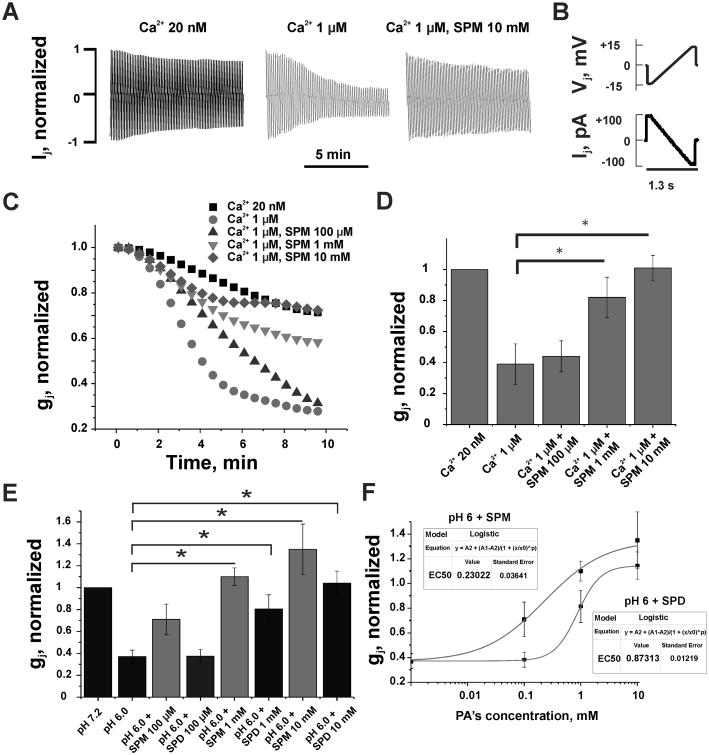
Figure 2. Polyamines prevented uncoupling of Novikoff cells induced by enhanced intracellular calcium or hydrogen concentrations.
(A) Examples of junctional currents (Ij) records in response to repeated (0.7 Hz) Vj ramps shown in (B). (C) Changes in the time course of averaged and normalized gj at different [Ca2+]i and spermine (SPM). An increase of [Ca2+]i from 20 nM to 1 μM accelerated gj decay (filled circles over filled squares). Addition of SPM at 0.1 mM (up triangles), 1 mM (down triangles) and 10 mM (diamonds) decelerated gj decay up to full recovery at 10 mM of SPM. (D) A summary of data demonstrating a lessening of Ca2+-induced uncoupling by SPM (p<0.05, n=7 in each group). (E) Averaged and normalized gj show that spermidine (SPD) diminish acidosis-mediated gj decay in a concentration dependent manner (p<0.05, n=6 in each group). Data shown in grey columns (SPM) demonstrate that SPD effect is lower than that of SPM at all three concentrations. (F) Dose-response relationships of normalized gj recovery under an influence of SPM and SPD. Curves are fits of a Logistic equation to the experimental data.

