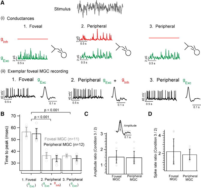
Figure 5. Inhibitory Synaptic Input Affects Gain but Not Kinetics of Peripheral Midget Ganglion Cell Outputs.

(A) Dynamic clamp experiment illustrating the three sets of conductances injected into foveal or peripheral MGCs. (1) Synaptic conductances were measured in response to white noise stimuli as in Figure 1C: 1. Foveal gExc 2. Peripheral gExc + gInh and 3. Peripheral gExc (2) Exemplar current-clamp recording from a foveal MGC where each of the three sets of conductances evokes corresponding spike responses. Exemplar STA from a foveal MGC for each of the three injected conductance sets.
(B) Bar graph comparing the time to peak of the STAs for each of the three conductance sets for foveal and peripheral MGCs. MGCs show faster kinetics with shorter time to peak when injected with peripheral synaptic conductances at the fovea (mean of 36 ± 3 ms for gExc + gInh and 36 ± 1 ms for gExc) and in the periphery (mean of 34 ± 3 ms for gExc + gInh and 34 ± 2 ms for gExc). However, when MGCs are injected with foveal conductances their response kinetics gets appreciably slower (mean of 57 ± 4 ms for foveal MGCs and mean of 55 ± 4 ms for peripheral MGCs).
(C) Ratio of the STA amplitude measured from MGCs without peripheral inhibitory conductances (set 3 in A) to those measured with peripheral inhibitory conductances (set 2 in A) yields a mean value of 1.5 ± 0.4 for both foveal and peripheral MGCs.
(D) The ratio of the mean spike rate yields a mean value of 2.2 ± 1.4 and 1.9 ± 0.5 for foveal and peripheral MGCs, respectively.
