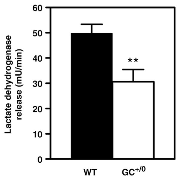
Fig. 1.

Index of membrane integrity in isolated hearts from control WTand GC+/0 mice. Data are means±SEM of 13–15 heart perfusion experiments. Values shown represent averages over the entire perfusion period. Lactate dehydrogenase release rates of control (WT) mice (solid bars) and hearts from mice overexpressing guanylate cyclase in a cardiomyocyte-specific manner (GC+/0) mice (open bars) were determined enzymatically by spectrophotometric method in effluent perfusates collected every 5 min. **P < 0.01 GC+/0 vs control WT mouse hearts.
