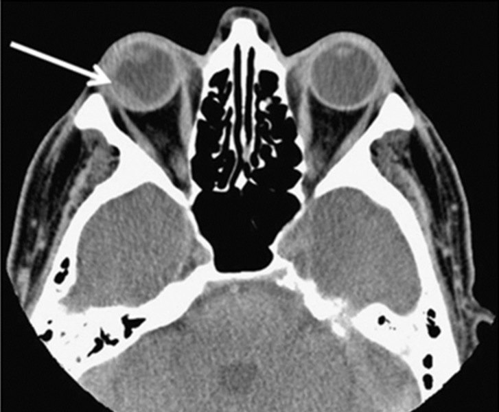
Figure 4. CT image demonstrating right ocular lymphoma lesion with extrascleral involvement.
CT scan showing a chorioretinal mass (arrow) in the right orbit, predominantly affecting the lateral and posterior parts of the globe, as well as transscleral extension laterally at the time of choroidal biopsy. (Reference: Mota, et al. [22]).

