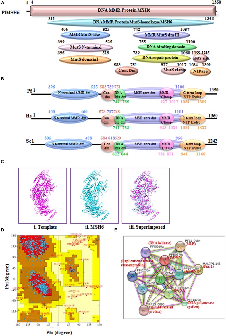
FIGURE 5.

(A) Detailed graphical organization of P. flaciparum MSH6 domains with their specific position in the protein. (B) Schematic comparison of aligned sequence of P. falciparum MSH6 with humans and Yeast; (C) Structural modeling of PfMSH6. (i) Template human MSH6 (PDB No. 2O8EB), (ii) PfMSH6 (iii) superimposed image (D) Ramachandaran Plot of PfMSH6 predicted structure. (E) Interacting proteins prediction of PfMSH6.
