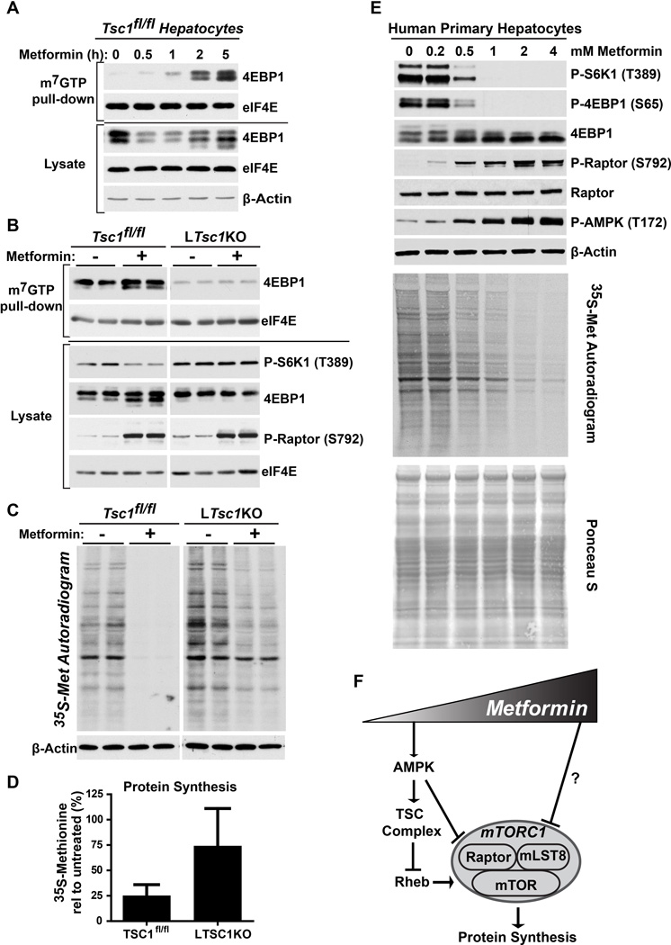
Figure 4. Metformin inhibits protein synthesis in hepatocytes through the TSC complex.
(A) Primary hepatocytes from TSC1fl/fl mice were treated with 1 mM metformin for the indicated times, and mRNA 5’-cap-binding complexes were isolated from cell lysates with m7GTP-agarose and analyzed by immunoblotting.
(B) Primary hepatocytes from TSC1fl/fl and LTsc1KO mice were treated with 1 mM metformin for 5 h and analyzed as in (A).
(C and D) Effects of metformin on hepatocyte protein synthesis. Primary hepatocytes from TSC1fl/fl and LTsc1KO mice were pretreated with insulin for 20 min followed by treatment in the presence or absence of 1 mM metformin for 5 h, with a pulse label of [35S]-methionine for the final 20 min. A representative autoradiogram from 5 independent experiments is shown. (D) Individual lanes from autoradiograms were quantified by densitometry and are graphed as mean ± SEM % incorporation relative to untreated cells (n=5 independent experiments). *P < 0.05 by Student’s two-tailed t-test.
(E) Primary human hepatocytes were treated with the indicated doses of metformin for 5 h and, for assaying protein synthesis, were radiolabeled as in (C). Signaling was assessed by immunoblots, protein synthesis by autoradiogram, and total protein for this assay by Ponceau S staining.
(F) Model of dose-dependent, differential regulation of mTORC1 in response to increasing metformin concentrations.

