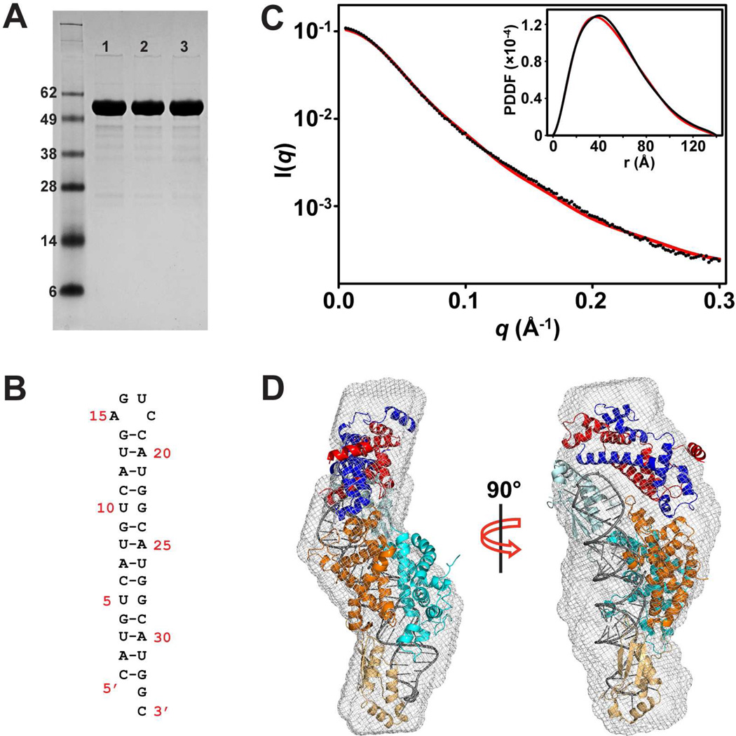
Figure 2.
Solution structure of Rnt1p post-cleavage complex by SAXS. (A) SDS-PAGE analysis of the Rnt1p protein used in the SAXS experiment. Lane 1 is apo-Rnt1p before the SAXS experiment. Lane 2 is apo-Rnt1p after the SAXS experiment. Lane 3 is the Rnt1p post-cleavage complex after the SAXS experiment. (B) Sequence and secondary structure of the product RNA when self-annealed. (C) Overlay of experimental scattering profiles (black) with back-calculated scattering profiles (red) for the complex. The insert shows the overlay of the structure (red) with experimental (black) PDDFs. (D) Shown in two views, crystal structure of the post-cleavage complex of Rnt1p (PDB entry 4OOG, excluding the extra NTD dimer) in the ab initio SAXS envelope. The protein domains and RNA are color coded as in Figure 1C.

