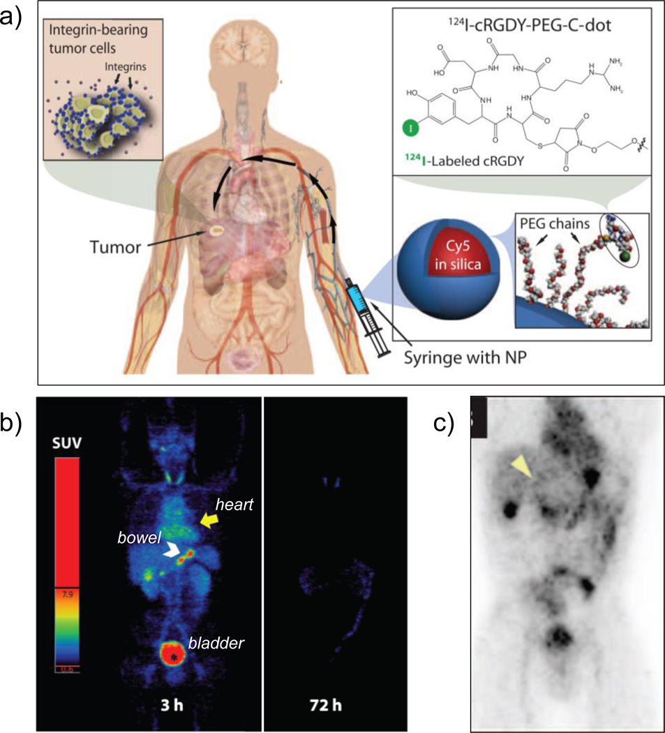
Figure 2.
(a) Schematic of the IND-approved hybrid (PET-optical) imaging nanoparticles (C dots). The core-contains Cy5 dye and the surface was attached with poly(ethylene glycol) (PEG) chains, integring αvβ3-binding cRGDY peptide ligands and 124I radiolabel. (b) Maximum intensity PET projections, 3 and 72 h after intravenous injection of 124I-cRGDY–PEG–C dots. (c) Coronal PET image 4 h p.i., demarcating the peripheral aspect of the tumor (arrowhead) and other major organs of nanoparticle uptake (bladder, gastro-intestina tract, gall bladder and heart). Adapted with permission from [36]. Copyright by American Association for the Advancement of Science.

