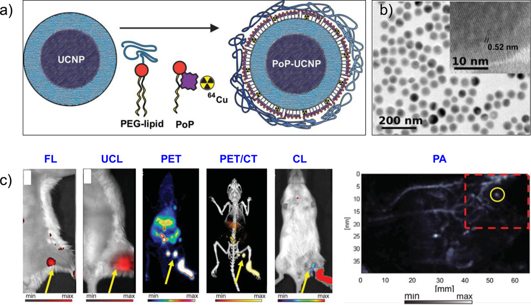
Figure 6.
(a) Schematic diagram of the PoP-UCNP structure. Core-shell UCNPs were transferred to the aqueous phase by lipid coating with PEG-lipid and porphyrin-phospholipid (PoP) followed by seamless intrinsic radiolabeling with 64Cu. (b) TEM image of PoP-UCNP, with the inset showing the crystalline and core-shell nanostructure of UCNP. (c) Hexamodal in vivo lymphatic imaging using PoP-UCNPs in mice via fluorescence imaging (FL), upconversion luminescence (UCL) imaging, PET, PET/CT, Cerenkov luminescence (CL) imaging, and photoaccoustic (PA) imaging. Reproduced with permission from [140]. Copyright by John Wiley and Sons.

