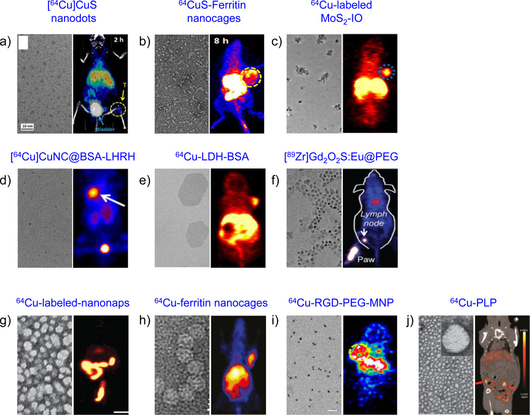
Figure 9.
Representative examples of other inorganic (a–f) and organic (g–j) nanomaterials employed for PET-guided cancer theranostics. Left panel: TEM images, right panel: in vivo PET images. (a) Copper sulfide nanodots; PET/CT image depicts 4T1 tumor uptake (yellow arrow) and dominant renal clearance (blue arrow; bladder) after i.v. injection of 5.6 nm sized [64Cu]CuS nanodots. Reproduced with permission from [284]. (b) Biomimetic CuS-Ferritin nanocages. TEM image depicts a dark CuS core inside a ferritin cage and PET image shows U87MG tumors (yellow circle), 8 h after i.v. injection of 64CuS-Ferritin nanocages. Reproduced with permission from [309]. (c) Iron oxide decorated MoS2 nanosheets. TEM image shows double-PEGylated MoS2-IO and PET image shows enhanced EPR-mediated uptake of intrinsically 64Cu-labeled MoS2-IO in 4T1 tumor-bearing mice (blue circle), 24 h p.i. Reproduced with permission from [286]. (d) High resolution TEM image of ultrasmall BSA-coated Cu nanoclusters (CuNCBSA). PET image depicts LHRH peptide-aided uptake of [64Cu]CuNCBSA-LHRH in orthotopic A549 lung tumor bearing mice (white arrow). Reproduced with permission from [288]. Copyright of American Chemical Society. (e) Layered double hydroxide (LDH) nanoparticles. Coronal PET image 16 h p.i. of 64Cu-LDH-BSA shows enhanced 4T1 tumor uptake. Reproduced with permission from [293]. Copyright of Nature Publishing Group. (f) TEM image shows Gd2O2S:Eu nanoparticles before PEGylation. PET-based lymph node mapping shows rapid delineation of sentinel lymph nodes 0.5 h after injection of [89Zr]Gd2O2S:EuPEG nanoparticles. Reproduced with permission from [294]. Copyright of John Wiley and Sons. (g) TEM image of ~20 nm frozen naphthalocyanine micelles (nanonaps) and PET image of 64Cu-labeled-nanonaps, 3 h after oral gavage. Reproduced with permission from [300]. Copyright of Nature Publishing Group. (h) TEM image of hybrid ferritin nanocages, conjugated to RGD targeting moiety and Cy5.5 dye. PET images of cRGDyK targeting of U87MG tumors, 24 h after the administration of ferritin nanoprobes. Reproduced with permission from [306]. (i) TEM image of multimodal PEGylated melanin nanoparticles (MNP; 10.7 nm) and PET images of 64Cu-RGD-PEG-MNP in U87MG tumor-bearing mice, 24 h p.i. Reproduced with permission from [305] (j) TEM shows a core-shell spherical structure of porphylipoprotein (PLP). PET/CT image of mouse with ovarian cancer metastasis after 24 h intravenous injection of 64Cu-PLP (red arrow: tumor). Reproduced with permission from [299]. Copyright of American Chemical Society.

