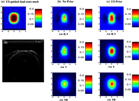
Fig. 2.
Reconstructed absorpstion map of the high contrast phantom target ( and ) at 780 nm at the 2.5-cm target depth. The maps at rest of the depths were not shown. (a) Reconstruction result of US-guided dual-zone mesh method. (b) Reconstruction result of NIRFAST with no prior using different . (c) NIRFAST with US prior. (d) Coregistered US B-scan image.

